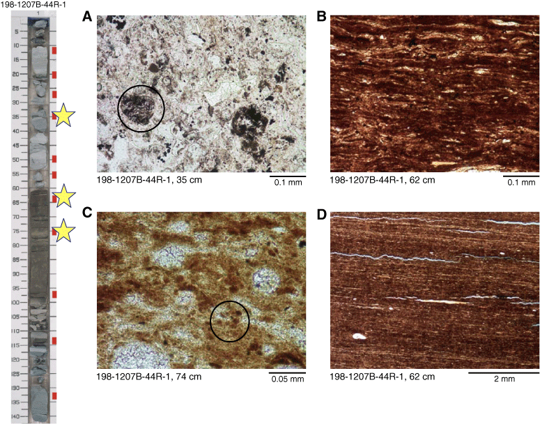
Figure F6. Pictured on the left is a core photograph with sample intervals and scale as in Figure F3. Stars highlight intervals corresponding to photomicrographs on the right (all in plane-polarized light). A. Main components in this tuff are blocky vitric fragments that are white because they have been altered to zeolites. Note rounded microlitic grain in left center. B. Laminated organic-rich shale is a close-up of the central portion of D. C. Small brown globular structures (example circled) can be seen in the matrix of this organic-rich radiolarian porcellanite. White circular features are altered radiolarians. D. Laminated structure in organic-rich interval exhibits blue, epoxy-filled fractures that are products of sample shrinkage during thin section preparation. White streak in center and blob in lower left are phosphatic fish debris.


![]()