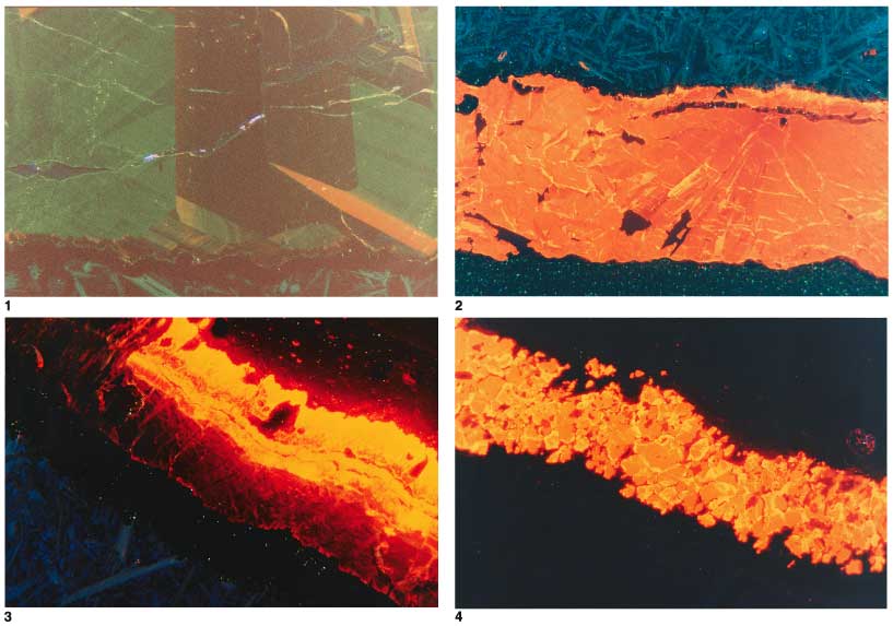
Plate 1. Photomicrographs of carbonate veins from Leg 168 basalt specimens, taken using cathodoluminescence (CL). 1. Blocky aragonite vein with basalt host rock visible along bottom edge. The aragonite is optically clear and homogeneous; however, the CL image reveals complex zones of varying CL color and intensity. Purple luminescence is from polishing compound (alumina?) stuck in epoxy that fills a microcrack through the section. Sample 168-1027C-5R-4, Piece 9. 2. Blocky and radial-fibrous calcite vein with basalt host visible along top. Although relatively homogeneous, bright areas may indicate zones of variable trace element concentrations. Sample 168-1027B-61X-CC, Piece 28. 3. Zoned vein with cross-fiber calcite in the middle (bright CL) and blocky aragonite (dark CL) at the edge. Host rock is visible at lower left. Sample 168-1032A-12R-1, Piece 1. 4. Blocky and granular calcite vein with zoned CL intensity. The outside edge of most calcite crystals have brighter CL, indicating more luminescence activators in the zone. Sample 168-1032A-12R-1, Piece 1. Click on image for close-up.
![]()