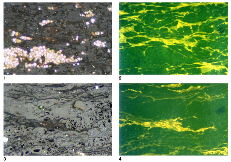
Plate P5.
Cell walls transitional to suberinite. 1.
T5809; Hole 1109D; 387.86 mbsf. Telovitrinite with fluorescing cell walls and a
prominent pyrite lens. Some of the fluorescing tissues are similar to those in
Plates P3 and P4
but some is similar to suberinite (reflected light; field width = 0.22 mm;
vitrinite reflectance [cell contents] = 0.31%). 2.
T5809; Hole 1109D; 387.86 mbsf. Same as figure 1, but in fluorescence mode. In
the upper right of the field the regular stacked structure normally associated
with suberinite is present, but in other parts of the field is it lacking, but
the cell walls show similar fluorescence properties (reflected light; field
width = 0.22 mm; vitrinite reflectance [cell contents] = 0.31%). 3.
T5814; Hole 1109D; 551.24 mbsf. Telovitrinite with abundant fluorescing cell
walls, some of which are suberinite. The part of the plant that has been
preserved is probably part of the periderm, and it appears that multiple
periderm layers are present in this wood type. The dark voids are similar in
appearance to resinite, but the fluorescence-mode plate shows that resinite is
not present and they represent open voids (reflected light; field width = 0.22
mm; mean vitrinite reflectance = 0.39%; suberinite reflectance = 0.18%). 4.
T5814; Hole 1109D; 551.24 mbsf. Same as figure 3, but in fluorescence mode. The
material most closely referable to suberinite is in the upper right of the
field, but this shows weaker fluorescence compared with the cell walls in the
center of the field. The structure and fluorescence of some of the layers toward
the base of the field are similar to those of cutinite (reflected light; field
width = 0.22 mm; vitrinite reflectance = 0.39%, suberinite reflectance = 0.18%).
Click on image or number to see enlargement.