![]() Figures F1-F50
Figures F1-F50
![]() Figures
F51-F100
Figures
F51-F100
![]() Figures
F101-F150
Figures
F101-F150
![]() Tables
T1-T27
Tables
T1-T27
F51. Unit 18 showing fine-scale flow lamination.
F52. Weakly developed alignment of plagioclase microlites and a single phenocryst in a silicified laminated rock.
F53. Anhedral former magnetite microphenocryst with remnant trellislike laths of magnetite faintly visible.
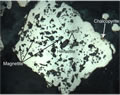
F54. Blotchy silicified rock with fine disseminated magnetite.
F55. Typical photomicrograph of strongly silicified sample showing a fine-grained intergrowth of granular quartz and phyllosilicates.
F56. Narrow anhydrite-pyrite vein with a typical cyclically banded alteration halo.
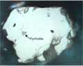
F57. Plagioclase microlites variably replaced by colorless illite and dirty brown pyrophyllite with white internal reflections.
F58. Plagioclase phenocryst completely replaced by fine-grained illite and possible halloysite.
F59. Quartz ± pyrite amygdules in a fine-grained quartz-illite groundmass.
F60. An apparently clastic interval.
F61. An apparently spherulitic unit, showing spherical domains in a cristobalite-clay matrix.
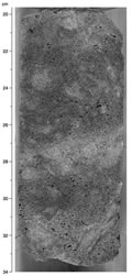
F62. Typical silicified unit from the lower sequence of Hole 1188F.
F63. Combined plane-polarized transmitted and reflected light photomicrographs of a "magnetite"-chlorite-quartz-pyrite filled vesicle.
F64. Dark magnetite-bearing halo around a white quartz-clay-pyrite-(anhydrite) vein.
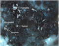
F65. Fine-grained green spinel hosted in quartz.
F66. Bladed hematite present as inclusions in quartz and as vesicle fill.
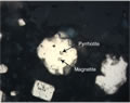
F67. Photomicrograph showing pyrite overgrowing a dark magnetite-bearing aggregate.
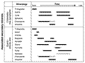
F68. Thin-section estimates of variation in abundance of significant minerals with depth for Holes 1188A and 1188F.
F69. Highly altered dacite fragments in a matrix of disseminated pyrite.
F70. Anhydrite-quartz-pyrite veins in highly altered dacite.
F71. Type 2 anhydrite-pyrite + cristobalite veins cut by a later anhydrite vein.
F72. Veins of anhydrite and pyrite together with disseminated pyrite in highly altered dacite.
F73. Same view as Figure F72 in reflected light.

F74. Subhedral pyrite containing vermicular inclusions of magnetite.
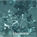
F75. Vermicular magnetite in quartz.
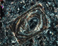
F76. Pyrite engulfing magnetite.
F77. Lamellate magnetite partially replaced by maghemite or hematite in an altered groundmass.
F78. Subhedral Fe-rich sphalerite precipitated on euhedral pyrite in a cavity.
F79. Vesicle filled with anhydrite, minor quartz, and pyrite.
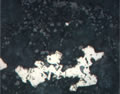
F80. Abundant inclusions in pyrite.
F81. Pyrrhotite inclusion and smaller magnetite inclusions in pyrite.
F82. Inclusions of pyrrhotite and magnetite in pyrite.
F83. Inclusion of hematite with a remnant of magnetite in pyrite.
F84. Hematite inclusions in pyrite.
F85. Chalcopyrite partially replacing pyrite.
F86. Ti magnetite with ilmenite exsolution laths.
F87. Ilmenite intergrown with magnetite surrounding spinel in a quartz matrix.
F88. Hematite flakes in quartz.
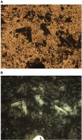
F89. Spinel crystals hosted in quartz.

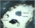
F90. Spinel, magnetite, ilmenite, and hematite.
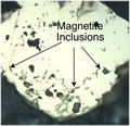
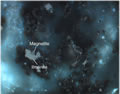
F91. Tiny magnetite inclusions in pyrite.
F92. Magnetite inclusions in pyrite having the same morphology as those in the neighboring quartz in the groundmass.
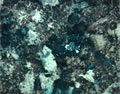
F93. Pyrite enclosing magnetite in a silicified rock.
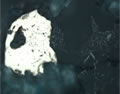
F94. Euhedral quartz overgrown by a second quartz containing inclusions of magnetite in turn overgrown by pyrite.
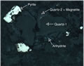
F95. Euhedral quartz-1 overgrown by magnetite and quartz-2 overgrown by pyrite.
F96. Paragenetic sequence.
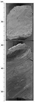
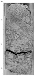
F97. Folded flow banding in pervasively altered volcanic rock, crosscut by silica-anhydrite-pyrite veins.
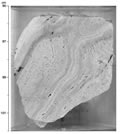
F98. Boudinlike disruption of flow lamination in pervasively altered volcanic rock.
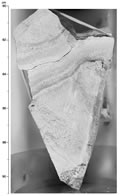
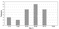
F99. Histogram of dips of volcanic layering, Hole 1188A.
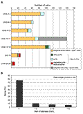
F100. Distribution of vein properties.
![]() Figures F1-F50
Figures F1-F50
![]() Figures F51-F100
Figures F51-F100
![]() Figures
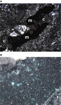
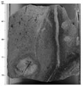
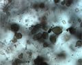
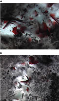
F101-F150
Figures
F101-F150
![]() Tables
T1-T27
Tables
T1-T27